Enthesis is the interface between tendon and bone in our body. Whilst it works perfectly well most of the time, if you rupture your enthesis, there is no way of healing it. I want to understand its nanostructure to find ways of fixing it.
Enthesis is an example of biomineralised tissues with a hierarchical structure. My goal is to find out the interplay between structure, function and formation. One of the key features of biomineralised tissues is that they have a hierarchical organisation that goes from the micrometre scale to the nanoscale and that is crucial for their mechanical properties. This has been the focus of my career: I started studying wood in my PhD, then bone and shell, to name a few examples.
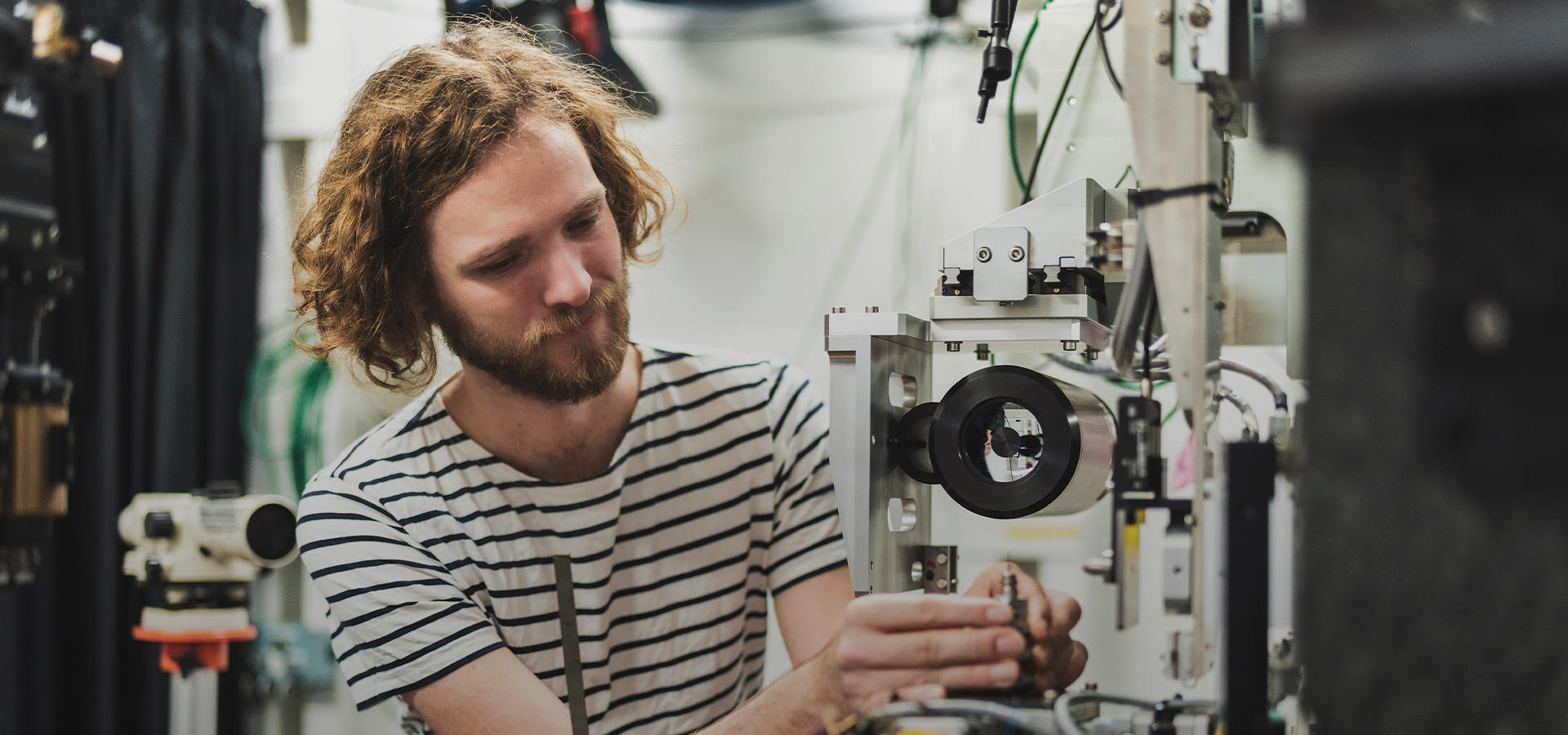
Today I am a CNRS researcher at the Institut Fresnel in Aix-Marseille University (France) and in the framework of my ERC grant, I will develop the new technique of texture tomography, a new 3D x-ray diffraction imaging method with support from beamlines ID13 and ID15A. Coupling texture tomography with micromechanical modelling will hopefully shed light onto the link between the hierarchical structure with the mechanical behaviour of the enthesis. It is clear that this interdisciplinary research is a requires input from various different sides and I am very happy to collaborate with the Institute des Sciences du Mouvement in Marseille on the biomechanics of the enthesis.
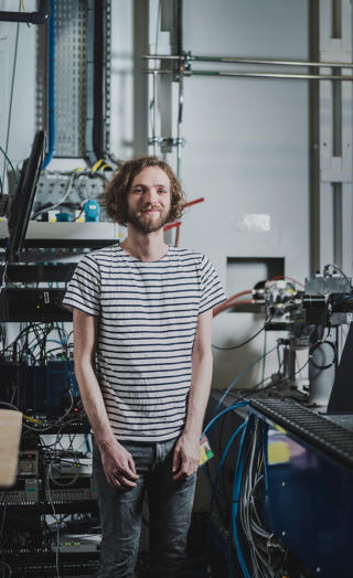
The ESRF has been a constant partner of my research over the last decade. I started my scientific career here. I came as a post-doc with lots of ideas and I had the chance to explore them thanks to the astonishing set of tools the facility offered me to do my research, including complementary beamlines and excellent support groups. The ESRF is also a great place to connect with experts and build up a network. This year I had the great honour of winning the Young Scientist Award.
Today the Extremely Brilliant Source is just what I need for my research. It feels as if someone had lifted the curtain and set the stage for a completely new kind of experiments. We can do previously unthinkable research, such as tracking how an organism mineralizes live or studying the mechanical behaviour of the enthesis in-situ.
I am now able to get sub 100 nm x-ray beam sizes with high flux at ID13 or to have a sub-micron beam at 100 keV at ID15a and this is really a game changer and the only way for me to achieve my scientific goals.
Nature still has many tricks up its sleeve. Understanding hierarchical structures like enthesis or molluscs and their properties will hopefully lead to making manufactured materials stronger and in a more eco-friendly manner.
TexTOM – Grant No. 101041871
X-ray texture tomography as a tool to enable, multiscale, in-situ imaging of the enthesis, a biological hinge between bone and tendon